

Post-doc position – team ACDC .
Images from our work.
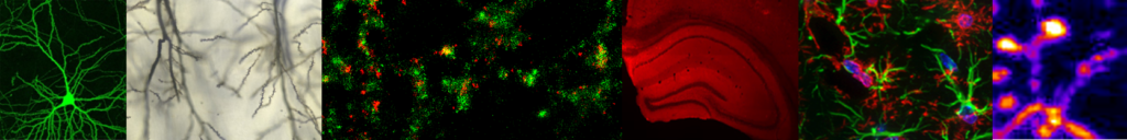
by Emilie Delaune. Team "Muscle formation, growth and repair"
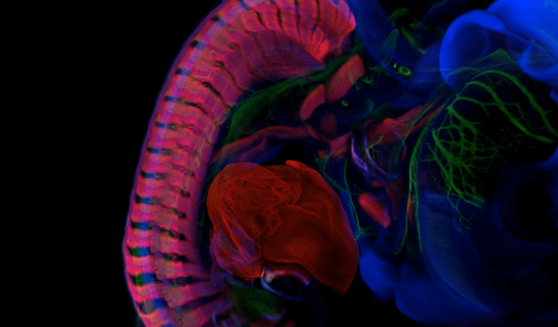
Chicken embryo at 5.5 days of development, clarified by the "3DISCO".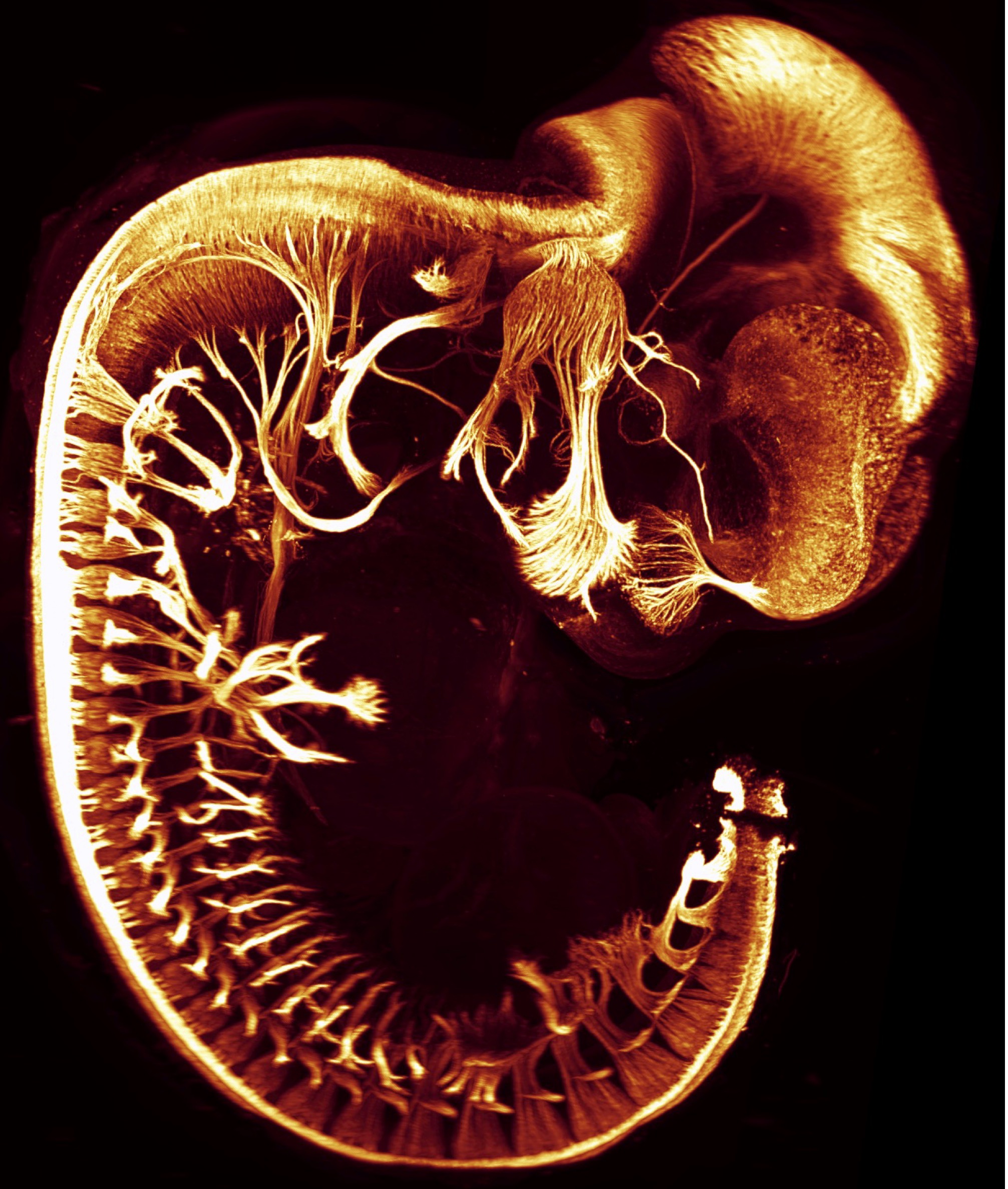
by V. Castellani. Team "Embryonic neuro-development and associated childhood diseases"
IF wholemount Tag1, embryon de souris KO Plex-M-Tyr , stade E11.5 (Tag1, Mouse embryo KO Plex-M-Tyr , stage E11.5).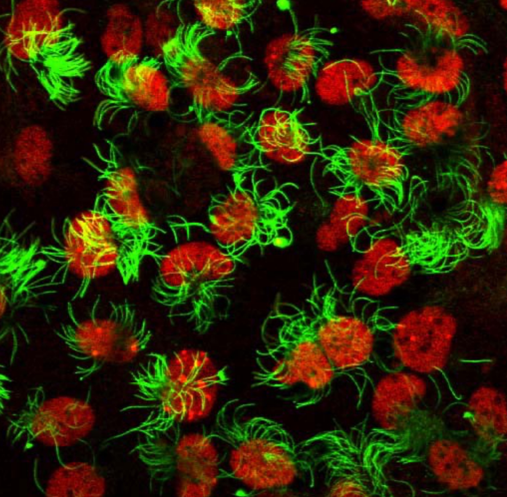
by B. Durand. Team "Cilia assembly and development"
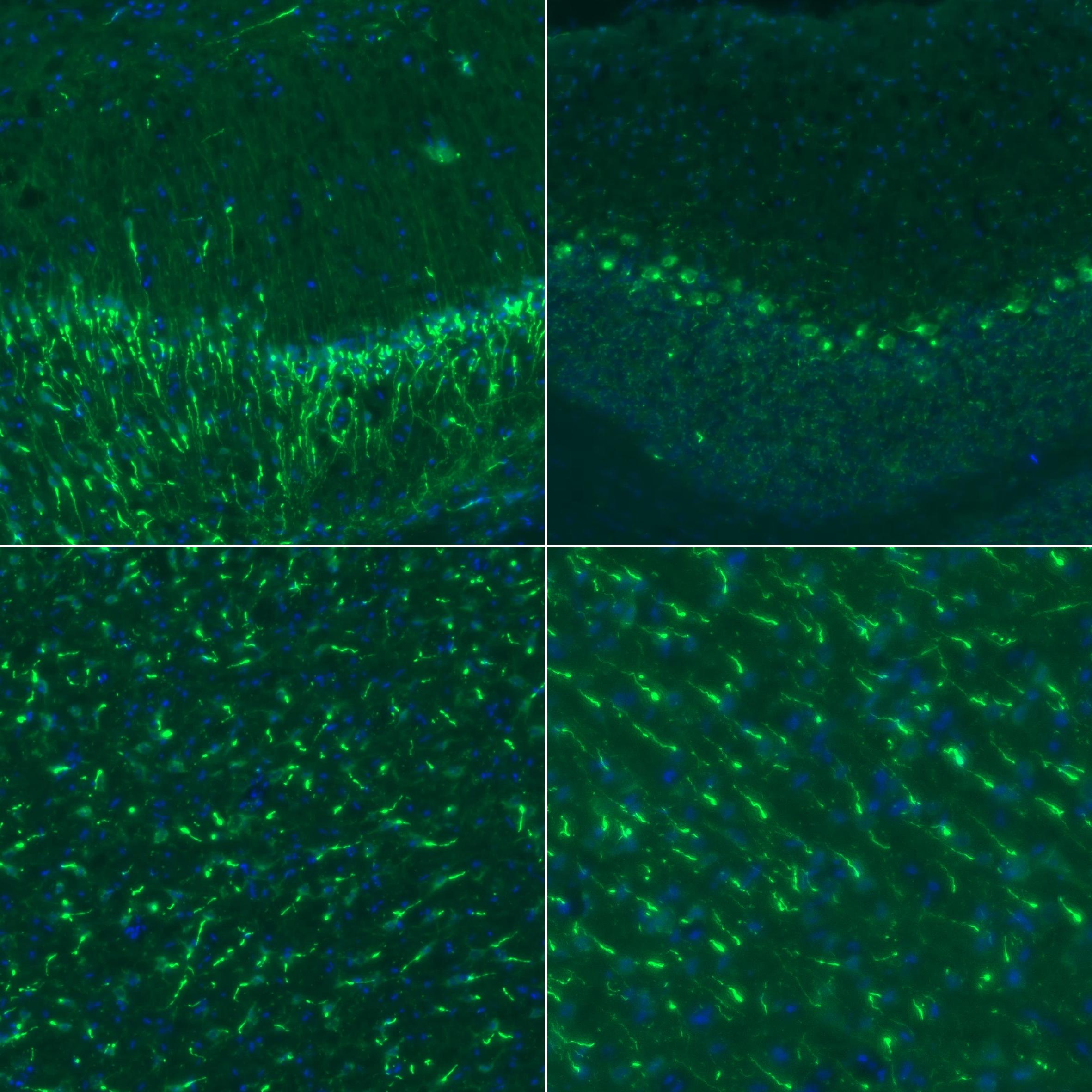
In vitro differentiated multiciliated mouse ependymal cells. Cilia are labelled in green (acetylated tubulin) and nuclei in red.
by V. Castellani. Team "Embryonic neuro-development and associated childhood diseases"
Triple marquage Ctip2, EdU Tbr1 sur coupe de cortex de souris à E13,5 (Ctip2 EdU Tbr1 Triple labelling on E13.5 mouse embryo neocortex).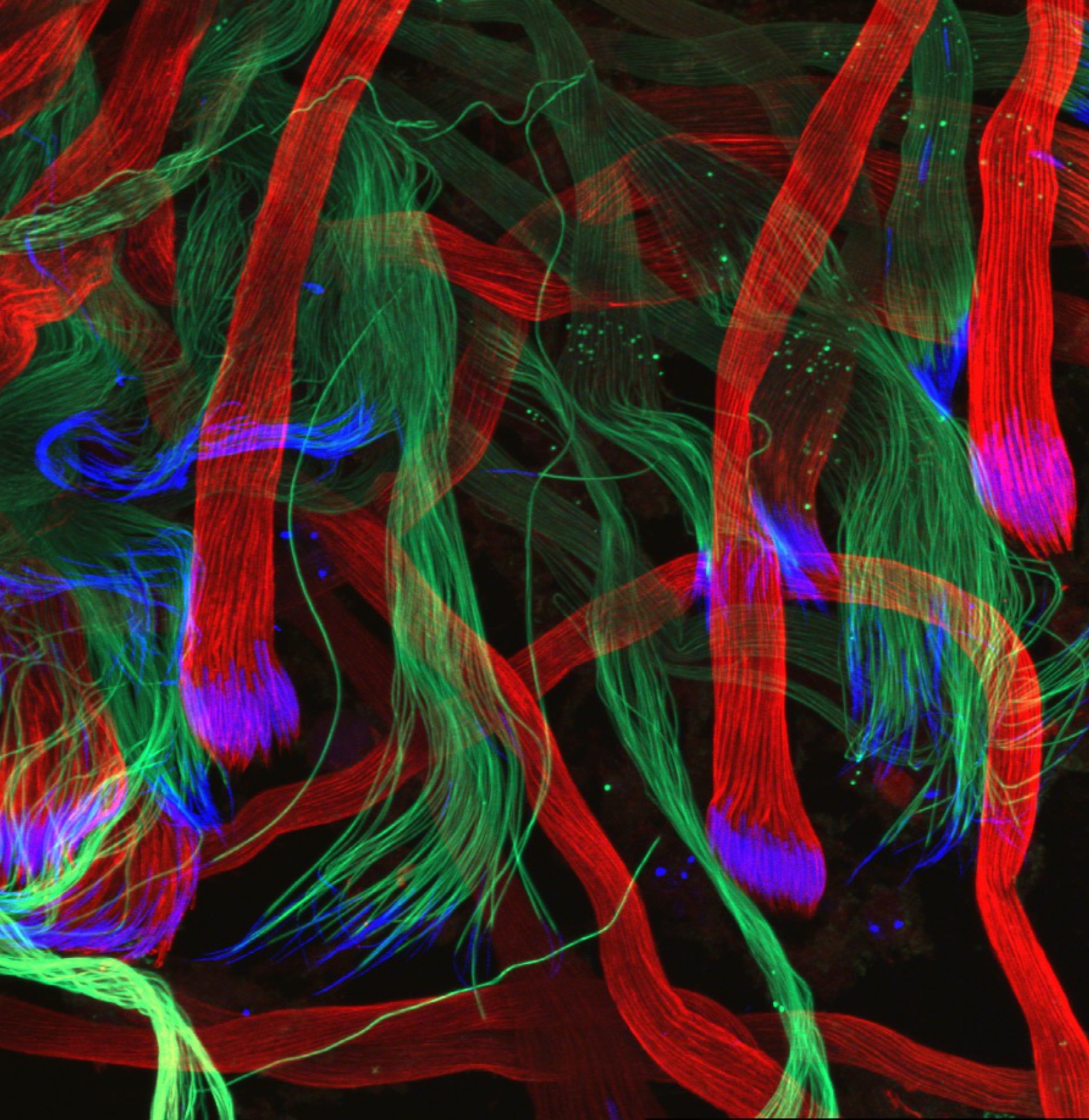
by B. Durand. Team "Cilia assembly and development"
Drosophila sperm cells at different maturation stages. Red: polyglutamylated tubulin; Green, polyglycylated tubulin; Blue: nuclei.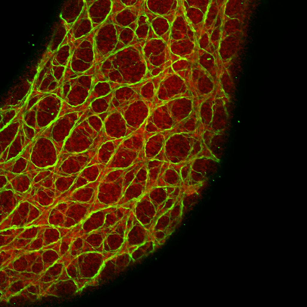
by V. Castellani. Team "Embryonic neuro-development and associated childhood diseases"
Système nerveux entérique d’embryon de poulet à E8 marqué par Neurofilament, et Phox2b (Enteric nervous system on E8 chick embryo, IF Neurofilament and PHOX2B).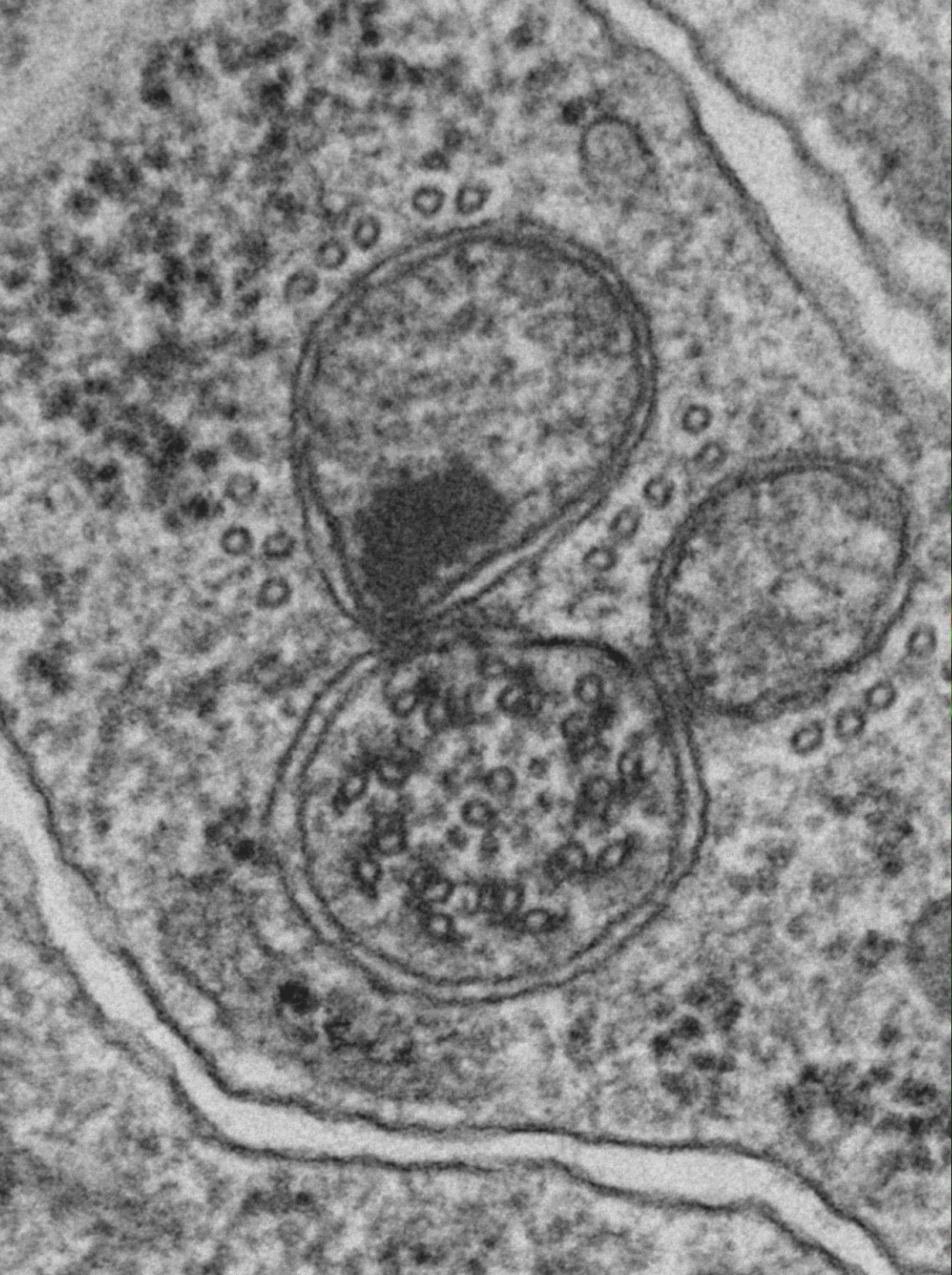
by B. Durand. Team "Cilia assembly and development"
Electron microscopy of Drosophila spermatids showing the axoneme and the two mitochondria derivatives.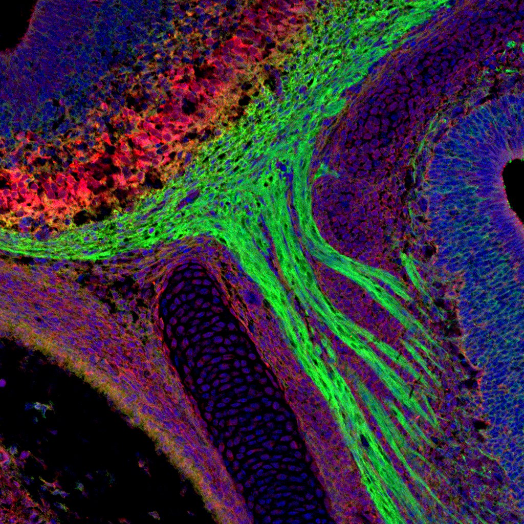
by V. Castellani. Team "Embryonic neuro-development and associated childhood diseases"
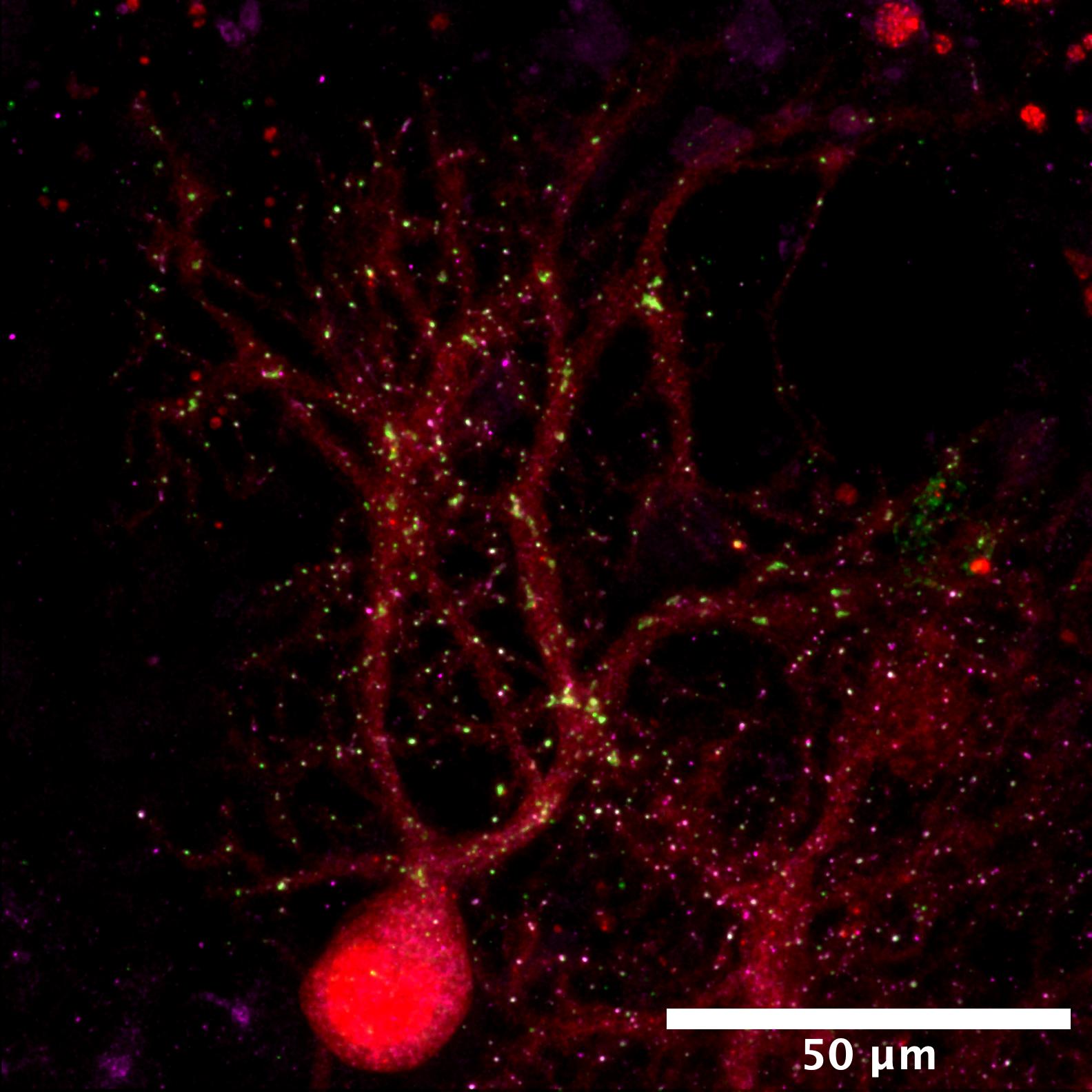
Nerfs olfactifs Plexin A1 vert et CHMP2B rouge (Olfactory nerves Plexin A1 green and CHMP2B red).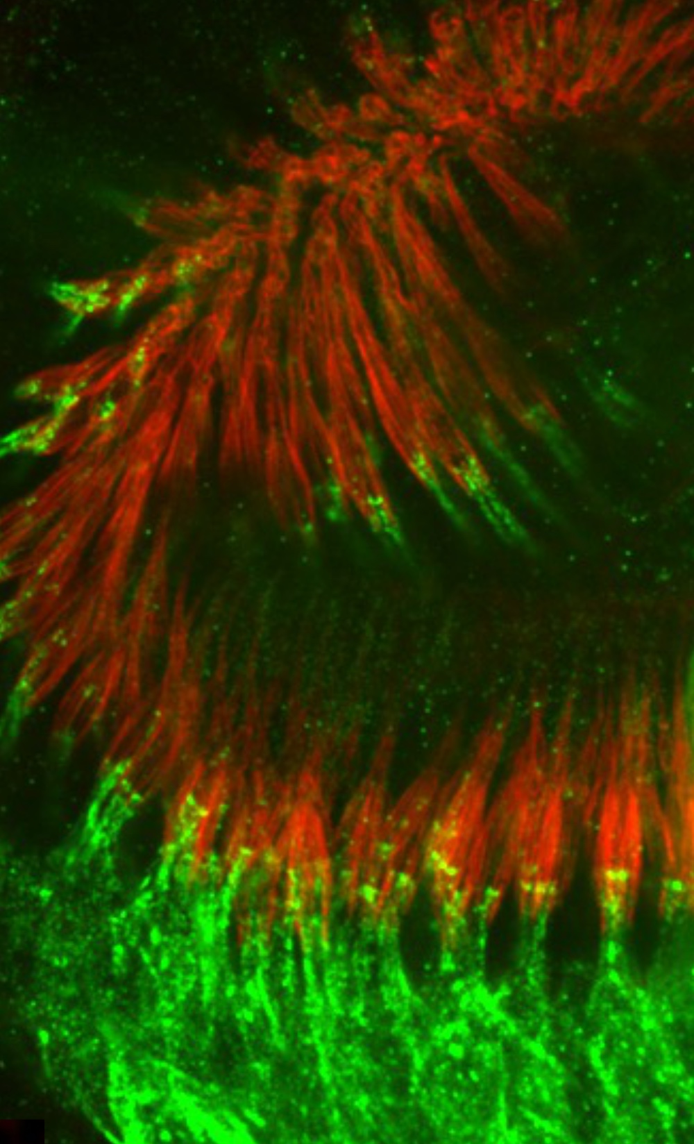
by B. Durand. Team "Cilia assembly and development"
Drosophila chordotonal neurons of the antennae: dendrites are labelled in green, actin associated strctures in red.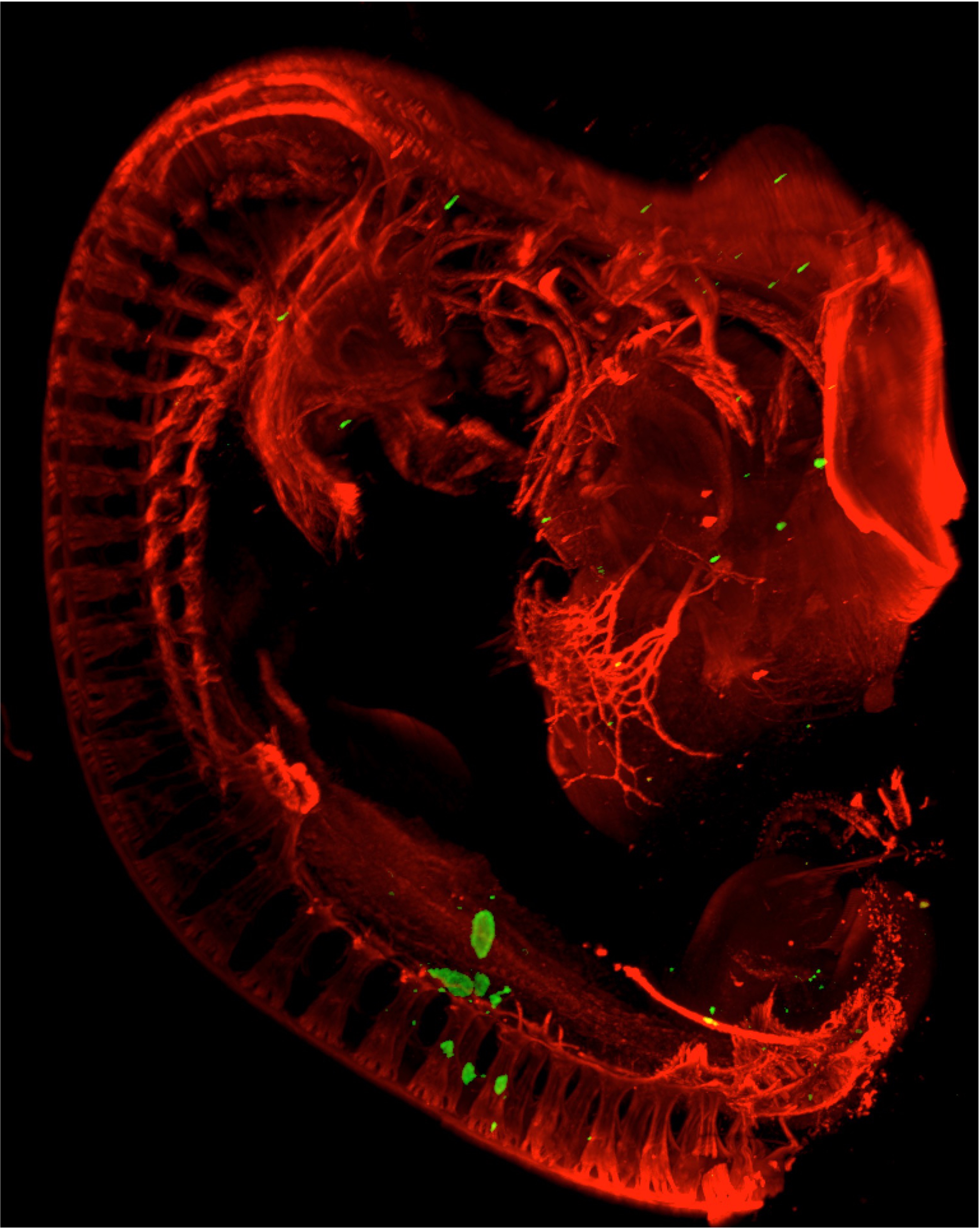
by V. Castellani. Team "Embryonic neuro-development and associated childhood diseases"
Tumeurs de neuroblastomes (vert) greffées dans un embryon de poulet, marquage GFP et HNK-1 à E5 (Neuroblastoma tumors (green) grafted in a chick embryo, GFP and HNK-1 staining at E5).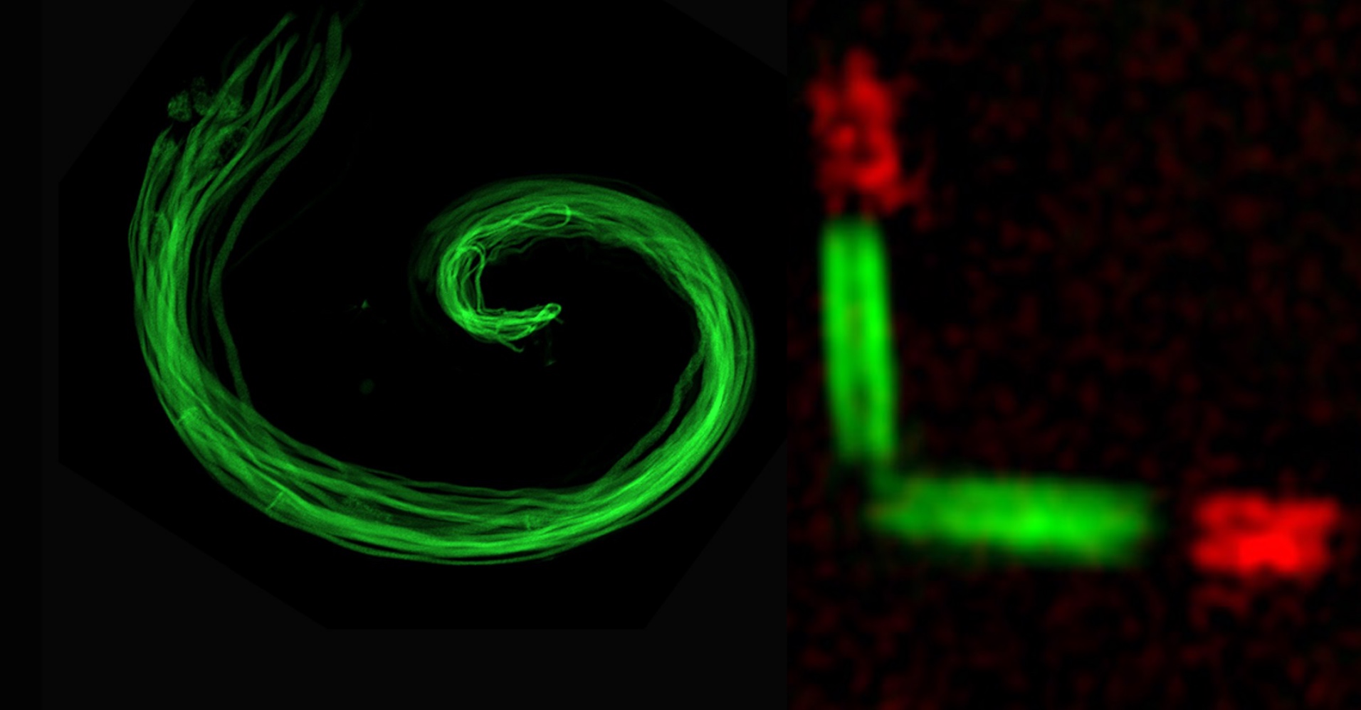
by B. Durand. Team "Cilia assembly and development"
A pair of Drosophila spermatocyte primary like cilia observed by 3D-SIM. Centriole in green, cilia in red.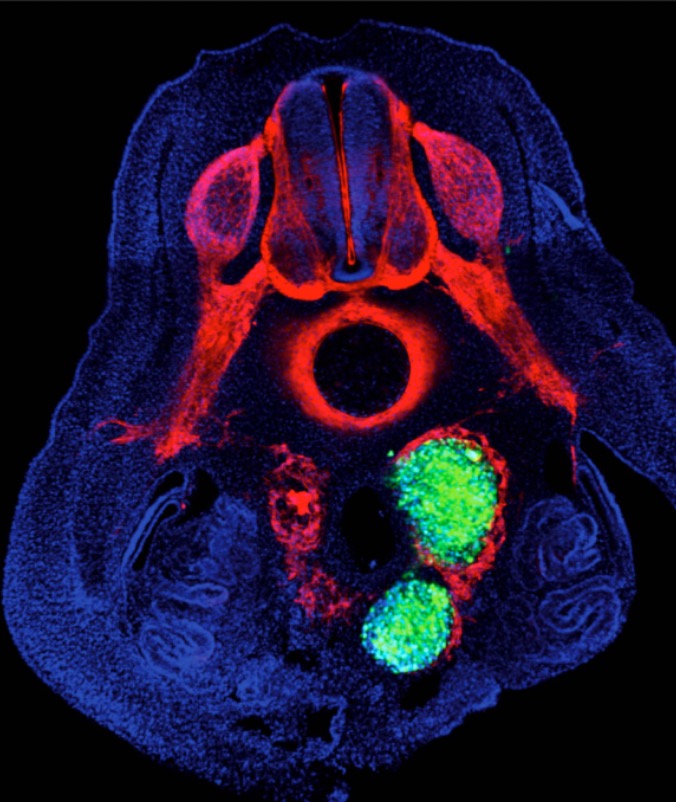
by V. Castellani. Team "Embryonic neuro-development and associated childhood diseases"
Tumeurs de neuroblastomes (vert) greffées dans un embryon de poulet, marquage GFP et HNK-1 à E5 (Neuroblastoma tumors (green) grafted in a chick embryo, GFP and HNK-1 staining at E5).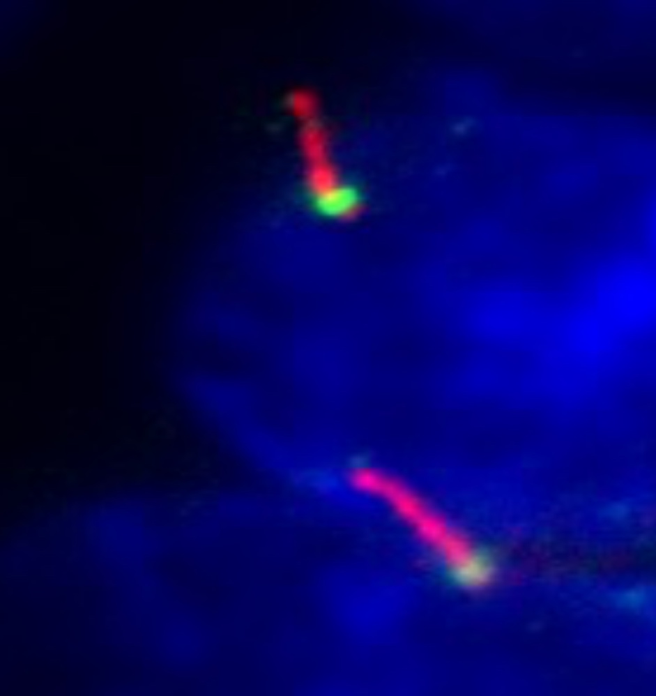
by B. Durand. Team "Cilia assembly and development"
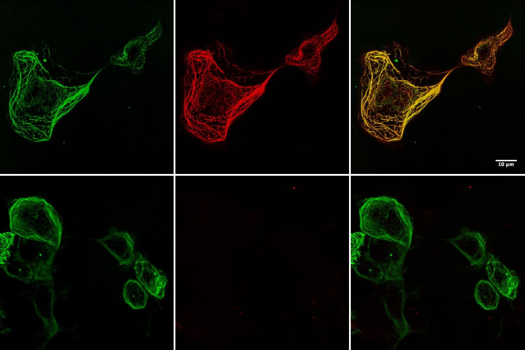
Mouse IMCD3 cells labelled for cilia (red), a basal body protein (CBY, green) and nuclei (blue).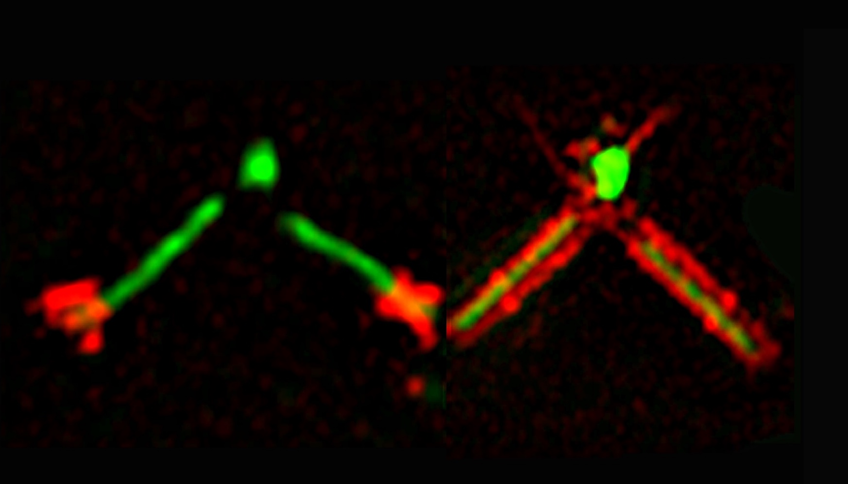
by B. Durand. Team "Cilia assembly and development"
Pairs of Drosophila spermatocyte centrioles observed by 3D-SIM. In green: Cep131; In red: Unc (right pair); Cby (left pair).

Post-doc position – team ACDC .
Phd or Engineer position – team SynatAc .

28 Nov - 11:00 – Salle Hermann
Julien DUPUIS, University of Bordeaux.
Rescuing pathological NMDA receptor hypofunction through synaptic trapping.
Direct inquiries about job opportunites to principal investigators. For other requests use the contact information below.